Cervical Cancer Stages
Cervical Cancer Stages was originally published by the National Cancer Institute.
A cancer stage describes the extent of cancer in the body, especially whether the cancer has spread from where it first formed to other parts of the body. It is important to know the stage of cervical cancer in order to plan the best treatment.
The International Federation of Gynecology and Obstetrics staging system is used for cervical cancer. Learn about tests and procedures used to determine the stage of cervical cancer.
Stage I cervical cancer
In stage I, cervical cancer has formed and is found in the cervix only. It is divided into stages IA and IB, based on the size of the tumor and the deepest point of tumor invasion.
Stage IA is subdivided based on the deepest point of tumor invasion.
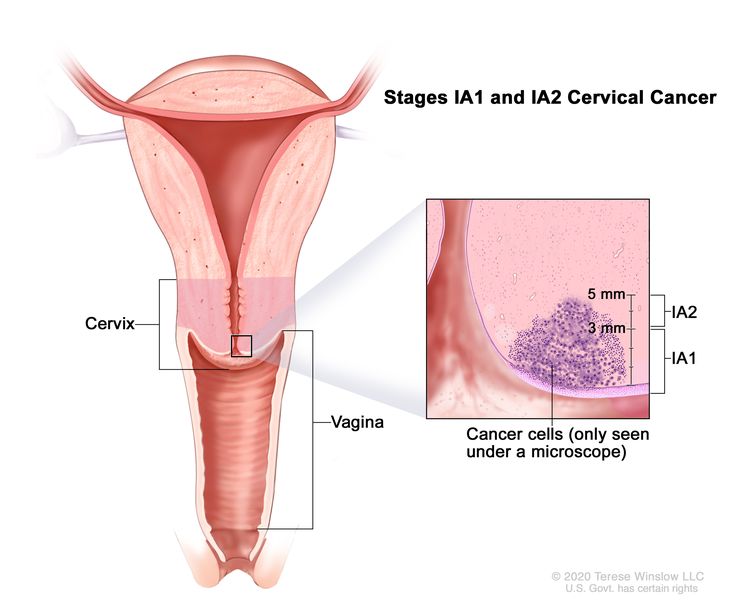
- Stage 1A1: A very small amount of cancer that can only be seen with a microscope is found in the tissues of the cervix. The deepest point of tumor invasion is 3 millimeters or less.
- Stage 1A2: A very small amount of cancer that can only be seen with a microscope is found in the tissues of the cervix. The deepest point of tumor invasion is more than 3 millimeters but not more than 5 millimeters.
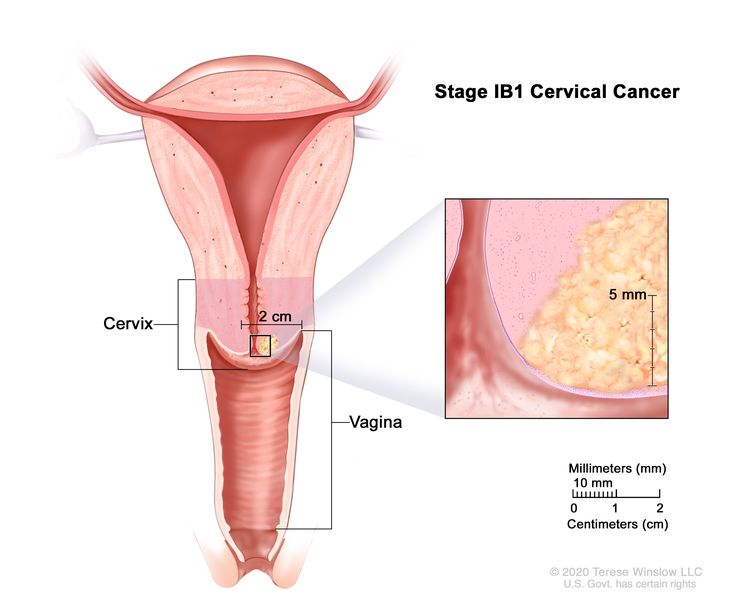
Stage IB is subdivided based on the size of the tumor and the deepest point of tumor invasion.
- Stage 1B1: The tumor is 2 centimeters or smaller and the deepest point of tumor invasion is more than 5 millimeters.
- Stage IB2: The tumor is larger than 2 centimeters but not larger than 4 centimeters.
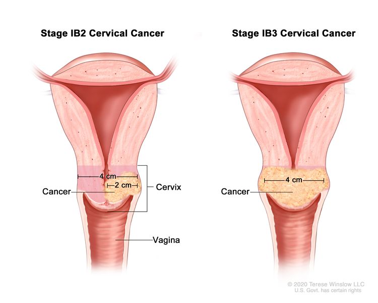
- Stage IB3: The tumor is larger than 4 centimeters.
Learn about treatment of stage I cervical cancer.
Learn about treatment of cervical cancer during pregnancy.
Stage II cervical cancer
In stage II, cervical cancer has spread to the upper two-thirds of the vagina or to the tissue around the uterus.
Stage II is subdivided based on how far the cancer has spread.
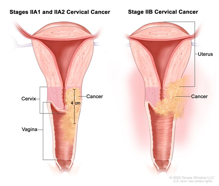
- Stage IIA: Cancer has spread from the cervix to the upper two-thirds of the vagina but has not spread to the tissue around the uterus. Stage IIA is further divided based on the size of the tumor:
- Stage IIA1: The tumor is 4 centimeters or smaller.
- Stage IIA2: The tumor is larger than 4 centimeters.
- Stage IIB: Cancer has spread from the cervix to the tissue around the uterus.
Learn about treatment of stage II cervical cancer.
Learn about treatment of cervical cancer during pregnancy.
Stage III cervical cancer
In stage III, cervical cancer has spread to the lower third of the vagina and/or to the pelvic wall, and/or has caused kidney problems, and/or involves lymph nodes.
Stage III is subdivided based on how far the cancer has spread.
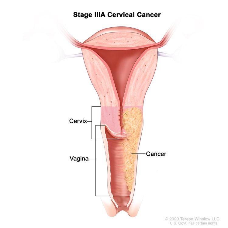
- Stage IIIA: Cancer has spread to the lower third of the vagina but has not spread to the pelvic wall.
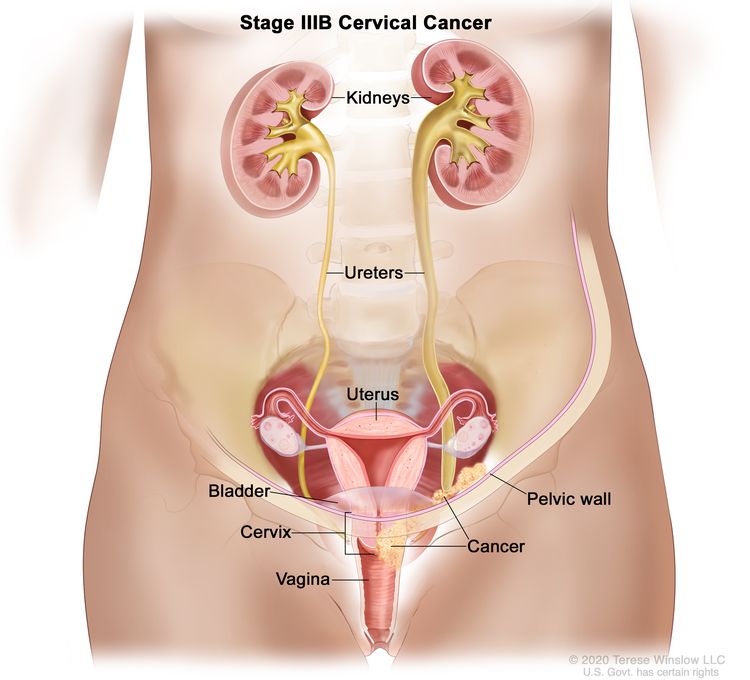
- Stage IIIB: Cancer has spread to the pelvic wall; and/or the tumor has become large enough to block one or both ureters or has caused one or both kidneys to get bigger or stop working.
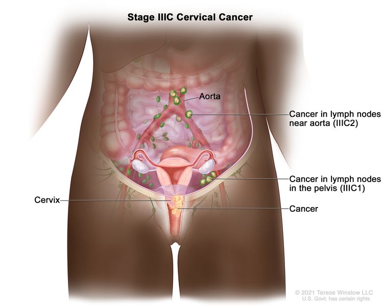
- Stage IIIC: Stage IIIC is divided into stages IIIC1 and IIIC2, based on the spread of cancer to the lymph nodes.
Learn about treatment of stage III cervical cancer.
Learn about treatment of cervical cancer during pregnancy.
Stage IV cervical cancer
In stage IV, cervical cancer has spread beyond the pelvis, or has spread to the lining of the bladder or rectum, or has spread to other parts of the body.
Stage IV is subdivided into stages IVA and IVB, based on where the cancer has spread.
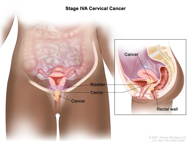
- Stage IVA: Cancer has spread to nearby pelvic organs, such as the bladder or rectum.
- Stage IVB: Cancer has spread to other parts of the body, such as the liver, lungs, bones, or distant lymph nodes

Stage IV is also called metastatic cancer. Metastatic cancer happens when cancer cells travel through the lymphatic system or blood and form tumors in other parts of the body. The metastatic tumor is the same type of cancer as the primary tumor. For example, if cervical cancer spreads to the lung, the cancer cells in the lung are actually cervical cancer cells. The disease is called metastatic cervical cancer, not lung cancer. Learn more in Metastatic Cancer: When Cancer Spreads.
Learn about treatment options for stage IV cervical cancer.
Learn about treatment of cervical cancer during pregnancy.
Recurrent cervical cancer
Recurrent cervical cancer is cancer that has recurred (come back) after it has been treated. The cancer may come back in the cervix or as metastatic tumors in other parts of the body. Tests will be done to help determine where the cancer has returned in your body, if it has spread, and how far. The type of treatment that you have for recurrent cervical cancer will depend on how far it has spread.
Learn about treatment of recurrent cervical cancer.
Learn more in Recurrent Cancer: When Cancer Comes Back.